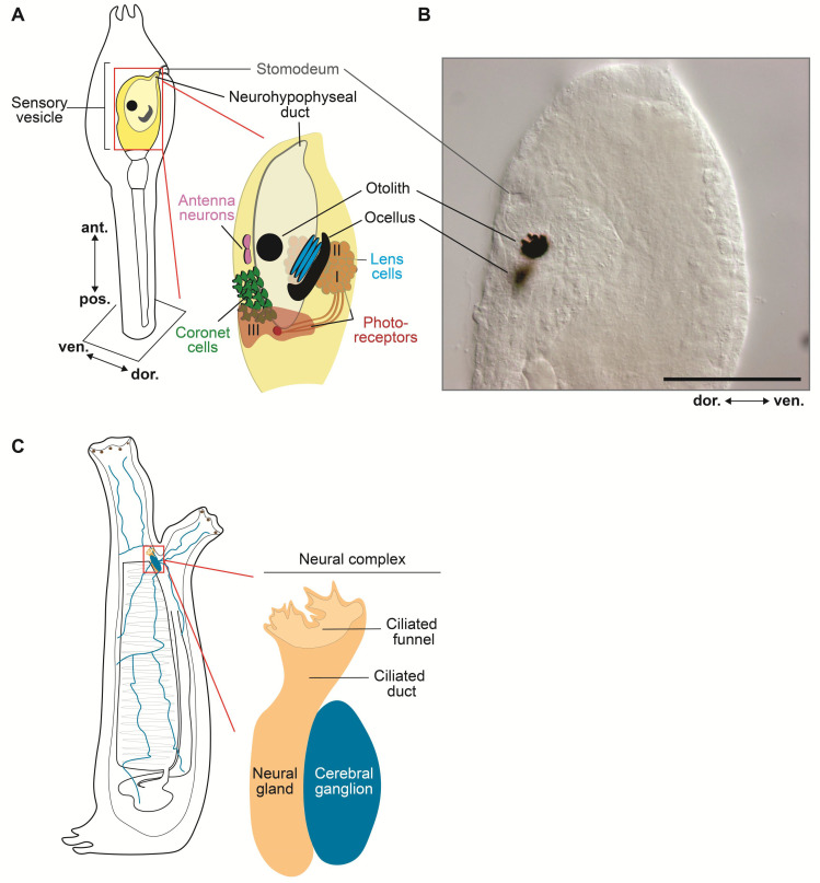
Figure 2.
Larval sensory vesicle and adult neural complex of Ciona. (A) Schematic view of the sensory vesicle, the ‘brain’ of the ascidian larva, its sensory organs, and the primordia of the hypophysis and stomodeum. On the right side of the sensory vesicle reside the pigmented ocellus and the associated lens cells and photoreceptors (group I and II), whereas the left side contains the otolith, antenna neurons, coronet cells, and photoreceptor cells (group III). (B) Microphotograph of the trunk of a Ciona larva, showing the developing stomodeum, the otolith, and the ocellus. Scale bar: 25 µm. (C) Schematic view of the sessile filter-feeder adult, highlighting the neural complex, located between the two siphons, and its components, the cerebral ganglion and the neural gland. Nerve fibers from the neural complex (blue) innervate multiple organs and tissues. Adapted from [48,85,86].

