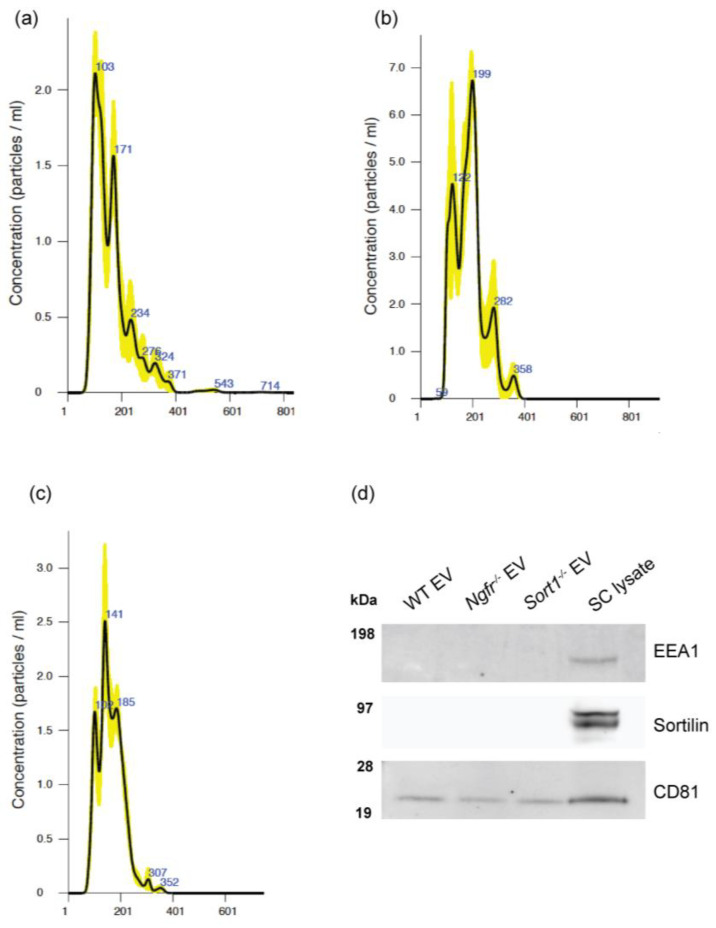
Figure 2.
Size distribution of vesicles secreted by the primary Schwann cells (SCs). Representative plots with nanoparticle tracking analysis results of extracellular vesicles (EVs) collected from 3 × T175 flasks from (a) WT, (b) Sort1−/− and (c) Ngfr−/− derived rat primary SCs. (d) Immunoblot showing CD81 expression in both the EV pellets and SC lysate, while EEA1 and sortilin were only found present in the SC lysate.

