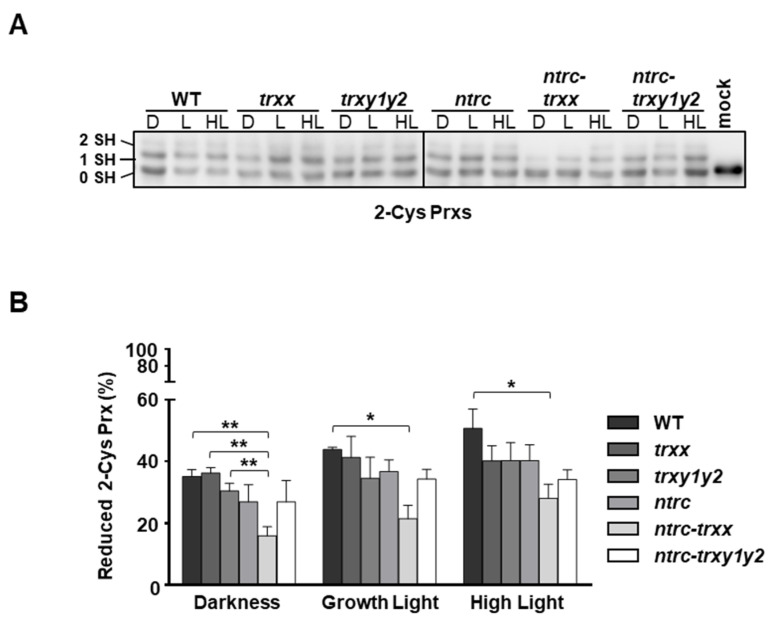
Figure 6.
In vivo redox state of 2-Cys Prxs in mutant plants devoid of Trxs y. Plants of the wild type and mutant lines, as indicated, were grown under long day conditions for 4 weeks. Whole rosettes were harvested at the end of the period of darkness (D) and then illuminated for 30 min with light intensities of 125 μE m−2 s−1 (growth light) or 450 μE m−2 s−1 (high light). (A) The in vivo redox state of 2-Cys Prxs was determined by alkylation using the thiol-labelling agent methylmaleimide-(polyethylene glycol)24 (MM(PEG)24). Mock indicates a control sample treated with alkylation buffer without MM(PEG)24. Protein extracts were subjected to SDS–PAGE under reducing conditions, transferred onto nitrocellulose filter and probed with an anti-2-Cys Prx antibody; 0 SH, 1 SH and 2 SH indicate reduction of none, one or the two cysteine residues, respectively, of 2-Cys Prxs. (B) Band intensities were quantified (GelAnalyzer), and the proportion of reduced protein was calculated as the ratio between the sums of half-reduced and fully reduced forms (1 SH + 2 SH) and the sum of oxidized (0 SH) and reduced forms (1 SH+2 SH). Each value is the mean of three independent experiments ± standard error (SE). Asterisks indicate significant differences between mean values (*, p < 0.05 and **, p < 0.01, Student’s t test).

