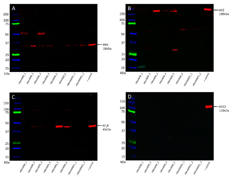
Figure 3.
Representative western blot images of head and neck metastatic malignant melanoma (HNmMM) tissue samples and HNmMM-derived primary cell line samples show varying expression levels of the components of the renin-angiotensin system. PRR ((A), ~28 kDa) was present in five out of six tissue samples and both HNmMM-derived primary cell lines at the expected size for the soluble form. ACE ((B), ~190 kDa) was only seen in three of the tissue samples. AT2R ((C), ~45 kDa) was present in four of the tissue samples, but none of the cell lines. ACE2 ((D), ~110 kDa) was not detected in any of the five tissue samples or three cell lines.

