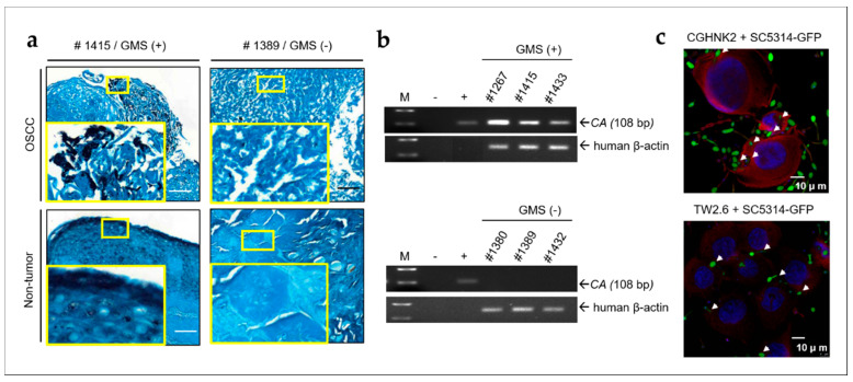
Figure 1.
CA (Candida albicans) is present in oral specimens from patients with OSCC (oral squamous cell carcinoma) and invaded CGHNK2 and TW2.6 cell lines. (a) GMS (Gomori methenamine silver) stain showing the presence of fungi in OSCC tumor and adjacent non-tumor tissues. Fungus was black stained. The number in each picture indicates the sample code. GMS-negative samples were shown as controls. Small and large yellow frames indicate the original and magnified areas, respectively. Bar, 100 μm. (b) Gel views of PCR products obtained with DNA extracted from OSCC biopsies. M, molecular weight marker (100-bp ladder); −, negative control; +, PCR carried out with DNA isolated from CA SC5314 as positive control. The size of the CA PCR product is indicated by an arrow (108 bp). Human β-actin PCR product used as internal control for each OSCC samples. (c) CA invade cultured human untransformed CGHNK2 and OSCC TW2.6 cell lines. Immunofluorescence confocal micrograph of CGHNK2 and TW2.6 cells after coculture with CA strain SC5314 expressing GFP (SC5314-GFP, MOI of 100) for 1 h. Invading intracellular CA (green) were confirmed by the single optical section through the host cells and indicated by white arrows. Fixed CGHNK2 and TW2.6 cells were stained with phalloidin-TRITC (red) to show filamentous actin. The nuclear counter stain is DAPI (4′,6-diamidino-2-phenylindole, blue). Bar, 10 μm.

