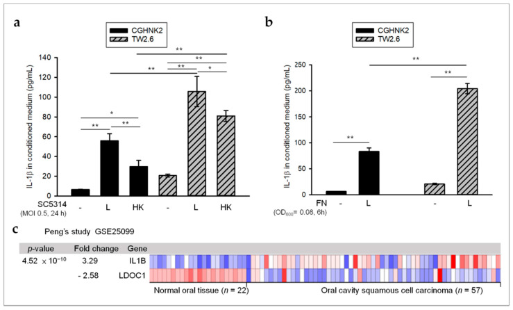
Figure 2.
Inverse relationship between LDOC1 and CA SC5314- or FN-induced IL-1β in OSCC. (a,b) LDOC1-deficient TW2.6 cells secrete more microbe-induced IL-1β as compared with LDOC1-expressing CGHNK2 cells. Cytometric bead array (CBA) analysis of IL-1β in conditioned medium of CGHNK2 and TW2.6 cell lines without microbes or with either live (L) or heat-killed (HK) CA SC5314 (MOI 0.5) (a) and L FN (OD600 = 0.08) (b) for 24 and 6 h, respectively. Results are presented as concentrations (pg/mL). Data are representative of at least three independent experiments (mean + S.D.), analyzed using the ANOVA test. * p < 0.05 and ** p < 0.01. (c) The mRNA expression profiles of LDOC1 and IL-1B in OSCC tumors (n = 57) and normal oral tissues (n = 22) were obtained from publicly available microarray data sets (GSE25099) [34] in Oncomine (https://www.oncomine.com).

