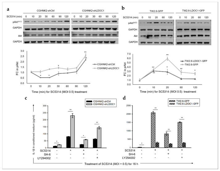
Figure 6.
The PI3K/Akt signaling pathway involved in CA-induced IL-1β production in LDOC1-deficient oral cells. (a,b) Effect of CA SC5314 on the phosphorylation of AktS473 in LDOC1-deficient or LDOC1-expressing oral cell lines. Cells cultured in a normal growth medium were treated with CA SC5314 (MOI = 0.5) for the times indicated. Equal amounts of whole-cell lysates were subjected to Western blotting analysis with antibodies specific for pAKTS473, Akt, and GAPDH. A representative blot is presented in the upper panels. As presented in the bottom panels, densitometric analyses were performed to quantify the fold change (FC) in the intensity of the pAKTS473 blots with untreated controls (time 0) set as 1. Data are expressed as mean ± SD (n = 2, biological duplicates). * p < 0.05, ** p < 0.01 vs. basal activation. (c,d) Levels of IL-1β induced by CA SC5314 were suppressed by inhibitors of Akt and PI3K (SH-6 and LY294002, respectively) in LDOC1-deficient CGHNK2-shLDOC1 (c) and TW2.6-GFP (d) cell lines. Concentrations of IL-1β in the conditioned medium of cells were measured by CBA after coculture with live CA SC5314 for 16 h, with or without pretreatment of SH-6 (2.5 µM and 5 µM for CGHNK2- and TW2.6-derived cell lines, respectively) or LY294002 (10 µM) for 6 h. Data are presented as mean ± SD (n = 3), analyzed using the ANOVA test. * p < 0.05 and ** p < 0.01.

