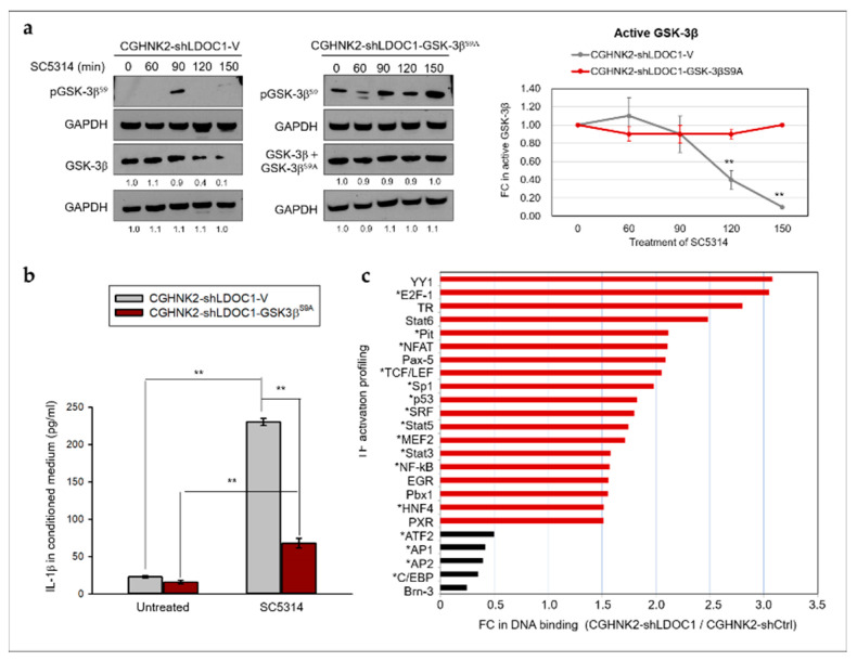
Figure 7.
GSK-3β participates in the enhanced IL-1β production in LDOC1-deficient CGHNK2-shLDOC1 cells after coculture with CA SC5314. (a) Western blotting analyses of the quantities of active GSK-3β, GSK-3βS9A, and inactive pGSK-3βS9 proteins in CGHNK2-shLDOC1-V (left panel) and CGHNK2-shLDOC1-GSK-3βS9A (center panel) cell lines. Densitometric analyses were performed to quantify the FC in the intensity of the active non-phosphorylated GSK-3β blots with untreated controls (time 0) set as 1 (right panel). Data are expressed as mean ± SD (n = 2, biological duplicates). ** p < 0.01 versus basal activation. (b) Concentrations of IL-1β in the conditioned medium were measured as described in the caption to Figure 4; three independent experiments were performed for IL-1β, each with three duplicates. Data are presented as mean ± SD (n = 3), analyzed using the ANOVA test. ** p < 0.01; (c) LDOC1 modulates immune-related TFs. Nuclear proteins isolated from CGHNK2-shCtrl and CGHNK2-shLDOC1 cells were subjected to a TF DNA-binding profiling assay. The fold change (FC) of intensities derived from TFII in CGHNK2-shLDOC1 versus CGHNK2-shCtrl was used for normalization. Experiments were performed according to the instructions provided by the manufacturer, as described in Materials and Methods. The bar charts represent the average FC values of two independent experiments.

