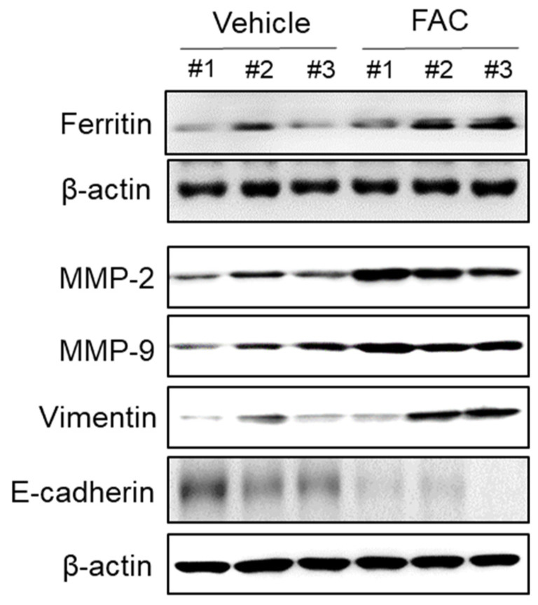
Figure 8.
Effect of iron on EMT and MMP-2/-9 expression in endometriotic lesions of mouse model. Female BALB/c mice were intraperitoneally administered with vehicle (0.05% CMC) and FAC (75 mg/kg) three times per week for 5 weeks. The endometriotic lesion tissue on peritoneum was lysed and the protein expressions of ferritin, MMP-2, MMP-9, vimentin, and E-cadherin were determined by Western blot analysis. β-Actin was used as a loading control. Each mouse tissue of the vehicle and FAC groups was marked with a number (#1–3).

