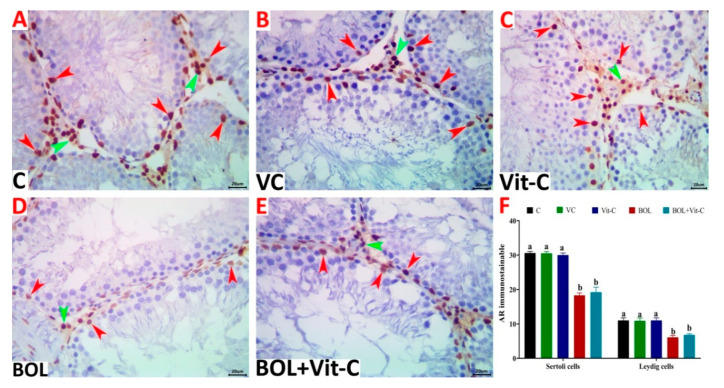
Figure 4.
(A–E) Representative photomicrograph of testicular tissue sections of androgen receptor (AR) immunoexpression showing a marked decrease in the numbers of AR immunostainable Sertoli (red arrowheads) and Leydig cells (green arrowheads) in control (A), vehicle control (VC) of sesame oil (B), vitamin C (Vit-C) treated (C), boldenone (BOL) treated (D), and Vit-C+BOL-treated rats (E). The scale bar is 20 microns. (F) Changes in numbers of Sertoli and Leydig cells in different experimental groups. Data are expressed as the mean ± SE (n = 8 replicates). Columns carrying different superscripts (a,b) are significantly different (one-way ANOVA) (p < 0.001).

