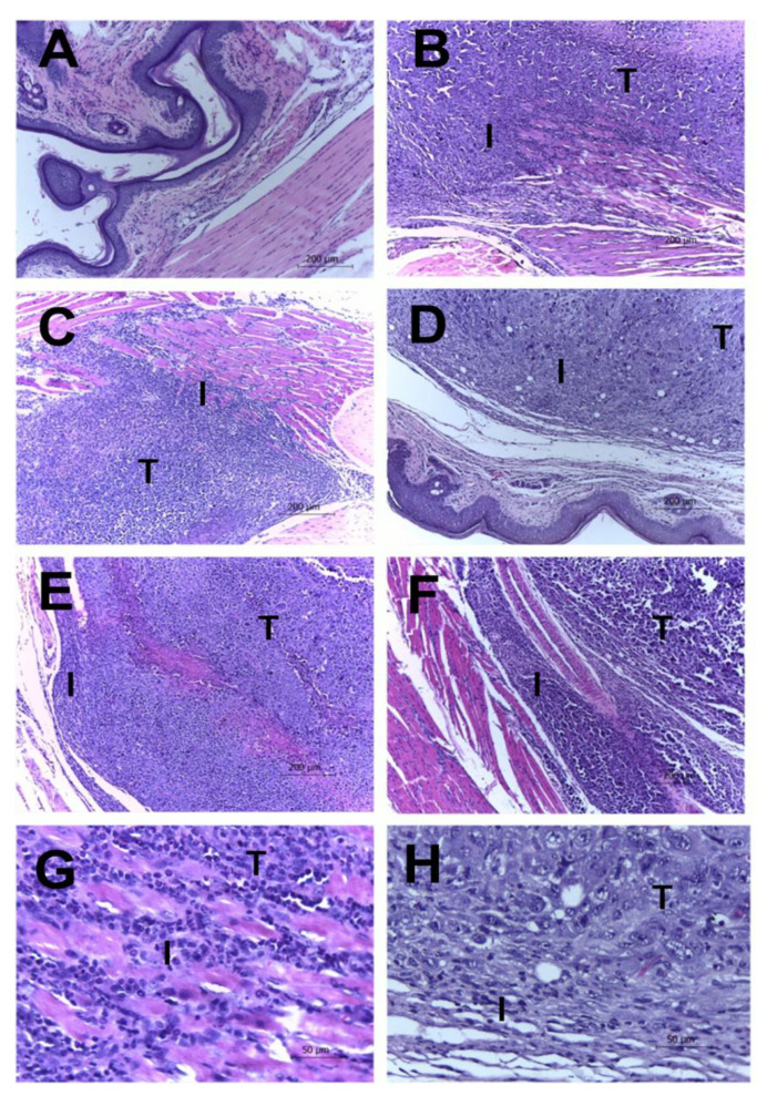
Figure 4.
Tumor and leukocyte infiltration in Ehrlich tumors with and without treatments. Leukocyte infiltrates in Ehrlich tumors. Swiss mice were inoculated in the paw with 2 × 10⁶ Ehrlich tumor cells and treated daily with EAE extract intraperitoneally. At the end of the fifteen days of treatment, the animals were euthanized, and their feet were amputated, weighed, and fixed. Histological sections were stained with Hematoxylin–Eosin. In the photos, it is possible to see the tumor cells (indicated by the letter T) and the inflammatory infiltrate (indicated by letter I) present in the paws of the Sham (A), CLT− (B), and CLT+ (C) groups (100× total magnification). The animals treated with the extract showed a decrease in the infiltrate and necrosis are EAE doses of EAE 4 (D), EAE 20 (E), and EAE 100 (F) (100× total magnification). The Sham group is shown in panel G, and the animals treated using the extract with a dose of EAE 100 are shown in panel H (400× total magnification).

