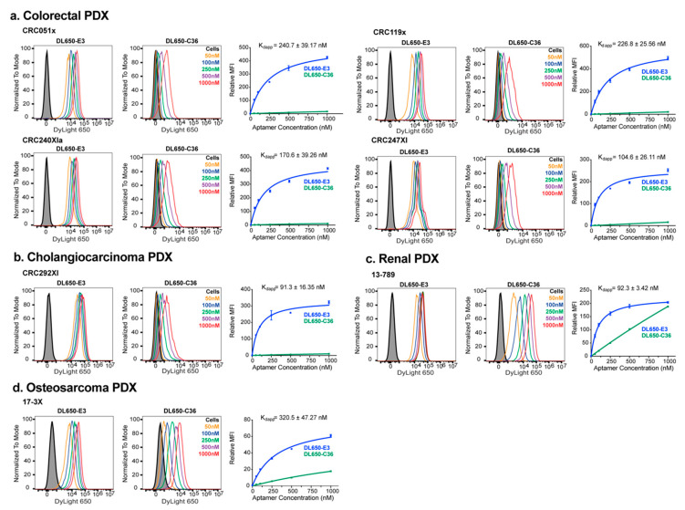
Figure 2.
The E3 aptamer targets broadly across different PDX-derived cancer cells. Cancer cells were incubated with DL650-E3 aptamer or DL650-C36 control aptamer and analyzed, as described in Figure 1. The binding curves are the combination of three independent experiments, with the median fluorescent intensity (MFI) of the aptamer-treated samples normalized to the median fluorescent intensity of the cells alone signal. Flow cytometry analysis of E3 targeting to (a) the colorectal PDX-derived cell lines CRC051x, CRC119x, CRC240XIa, and CRC247XI; (b) the cholangiocarcinoma PDX-derived cell line CRC292XI; (c) the renal cancer cell line 13-789; and (d) the osteosarcoma cancer cell line 17-3X.

