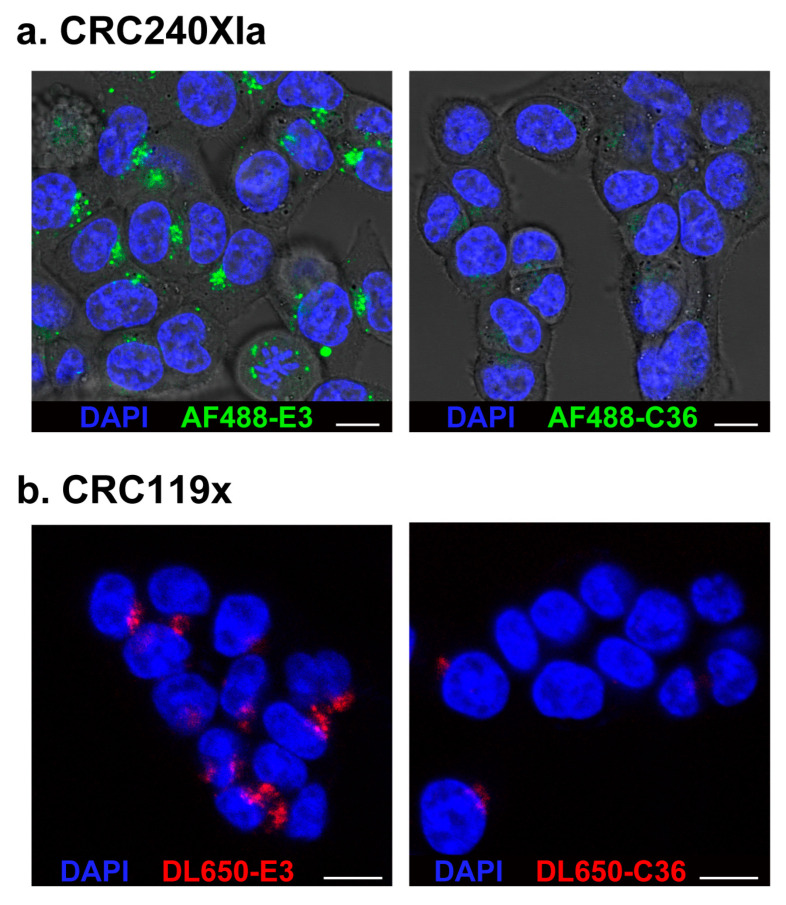
Figure 3.
Confocal microscopy confirms E3 aptamer internalization into PDX-derived cancer cells. Cells were treated for 1 h with (a) 10 μM of AF488-E3 aptamer or AF488-C36 control aptamer or (b) 1 μM of DL650-E3 aptamer or DL650-C36 control aptamer. After washing, Hoechst 33342 was added to all samples to stain the nuclei. Cells were imaged on a Leica SP5 inverted confocal microscope. (White scale bars: 10 μm).

