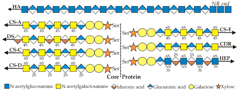
Figure 1.
Glycosaminoglycan diversity. The schematic representation of glycosaminoglycans (GAG) structures shows that heparin (Hep), dermatan sulfate (DS), chondroitin (CDR), chondroitin sulfates A (CS-A), C (CS-C), D (CS-D), and E (CS-E), but not HA, are synthesized onto core proteins (dashed line), which can be membrane-bound or soluble, via a common linker sequence (Galactose-Galactose-Xylose; two circles-star) attached to Ser. All GAGs are made as linear polymers with repeating disaccharide units containing either N-acetylglucosamine (GlcNAc, blue square) or N-acetylgalactosamine (GalNAc, yellow square) and either glucuronic acid (GlcU, blue diamond) or iduronic acid (IdU, brown diamond). Arrows denote the polymer growing end, to which new sugars are added. Sulfation occurs during biosynthesis and different cell types can create different negative charge patterns (unique molecular signatures) that are specifically recognized by other soluble or membrane proteins (e.g., Hyaluronic Acid Receptor for Endocytosis (HARE)). GAGs built on a core protein are attached at their reducing end, and new sugars are added at the nonreducing end. In contrast, HA is not attached to or built onto a core protein. Rather, HA synthesis is initiated de novo and extended by HA synthase until the UDP-HA chain is released, at which point it cannot be rebound and extended further [2]. Additionally different is that HA synthesis occurs at the reducing end, which is still attached to UDP. Thus, the first sugars assembled remain at the nonreducing end (NR end) and the linkages in this unique region are chitin [GlcNAc(β1,4)n] rather than HA. HA synthase initiates HA disaccharide synthesis only after first assembling a chitin-UDP (UDP-GlcNAc3-4) primer for starting HA synthesis [2]. The primer then remains as a chitin cap at the NR end [3].

