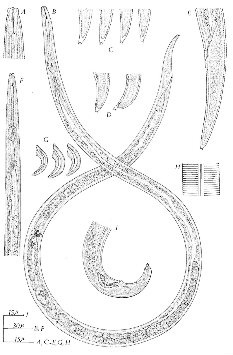
Figure 6.
Aphelenchoides ritzemabosi. (A) Female head; (B) female; (C) female tail ends; (D) male tail ends; (E) female tail; (G) spicules; (H) lateral field; (I) male tail region. (A, E, and F syntypes; B, C, and H Specimens from chrysanthemum, Stockholm; I Specimen from Buddleia leaf, Sussex, England) after Siddiqi [25]. Courtesy of Commonwealth Institute of Helminthology.

