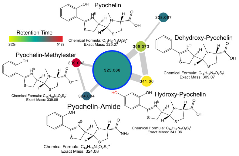
Figure 3.
Pyochelin diversity detected in clinical isolates of P. aeruginosa culture extracts. Structures, chemical formulas and exact masses of known and putative compounds are shown. The network nodes are colored by retention time according to the scale and sized by the number of spectra in the dataset. Edge width in the network is sized by the cosine score. Note that the hydroxyl highlighted in red is at an unknown position on the aromatic ring.

