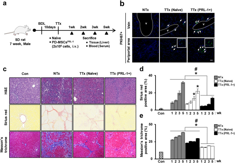
Fig. 5.
In vivo nonviral PD-MSCsPRL-1 alleviate liver fibrosis in a rat BDL model. a A schematic diagram describing the rat BDL model (NTx; n = 20, TTx naïve; n = 20, TTx PRL-1+; n = 20) as well as sham controls (Con; n = 5). b Engraftment of PKH67 (green)-labeled transplanted cells as injured rat liver by fluorescence microscopy at 1 week (arrow: PKH67+ signals) (green: PKH67; blue: DAPI). Scale bars = 100 μm. c Representative images of histopathological analysis in rat liver sections stained with H&E, Sirius red, and Masson’s trichrome after naïve and PD-MSCPRL-1 transplantation at 3 weeks. Scale bars = 100 μm. Quantification of d Sirius red- and e Masson’s trichrome-positive areas at 1, 2, 3, and 5 weeks in stained liver slides (n = 5). Data from each group are presented as the mean ± SD. *p < 0.05 versus NTx group; #p < 0.05 versus TTx naïve group. BDL, bile duct ligation; Con, control group; H&E, hematoxylin and eosin; NTx, nontransplantation group; PRL-1, phosphatase of regenerating liver-1; TTx, transplantation group

