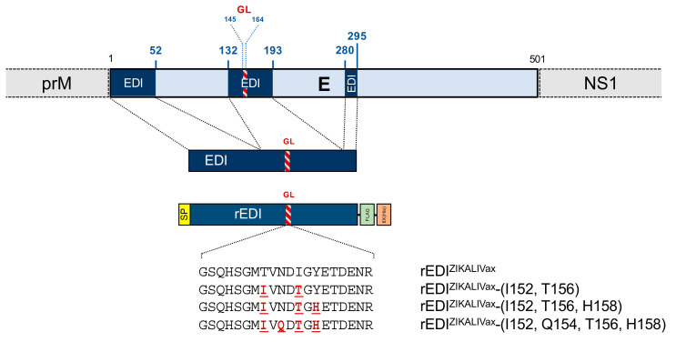
Figure 6.
Schematic representation of ZIKALIVax rEDI constructs. The organization of the E protein into ZIKV polyprotein with its three segments composing the EDI domain is shown on top. The EDI residues are numbered as those in the E protein of ZIKALIVax. The glycan loop (GL) sequence of E is shown as a red, hatched segment. The rEDIZIKALIVax sequence is preceded by a N-terminal heterologous signal peptide (SP) and followed by the spaced two C-terminal FLAG and 6x(His) tags in tandem. The rEDIZIKALIVax gene was inserted into plasmid vector pcDNA3. Directed mutagenesis was performed on rEDIZIKALIVax sequence to generate three mutants. The amino-acid substitutions introduced into rEDIZIKALIVax are indicated in red bold.

