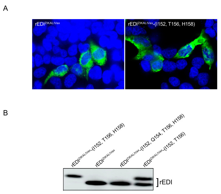
Figure 8.
Expression of rEDIZIKALIVax in HEK-293T cells. HEK-293T cells were transfected 24 h with plasmids pcDNA3 expressing rEDIZIKALIVax or its mutants. In (A), IF assay was performed on fixed and permeabilized cells with anti-6x(His) monoclonal antibody (mAb) as primary antibody. Alexa 488-conjugated anti-mouse IgG antibody as secondary antibody. The nuclei were stained with DAPI (blue). Immunostained cells were visualized with a fluorescent microscope. The same magnification of ×100 was used throughout. In (B), immunoblot was performed on RIPA lysates obtained from transfected cells expressing rEDIZIKALIVax or its mutants using anti-FLAG antibody.

