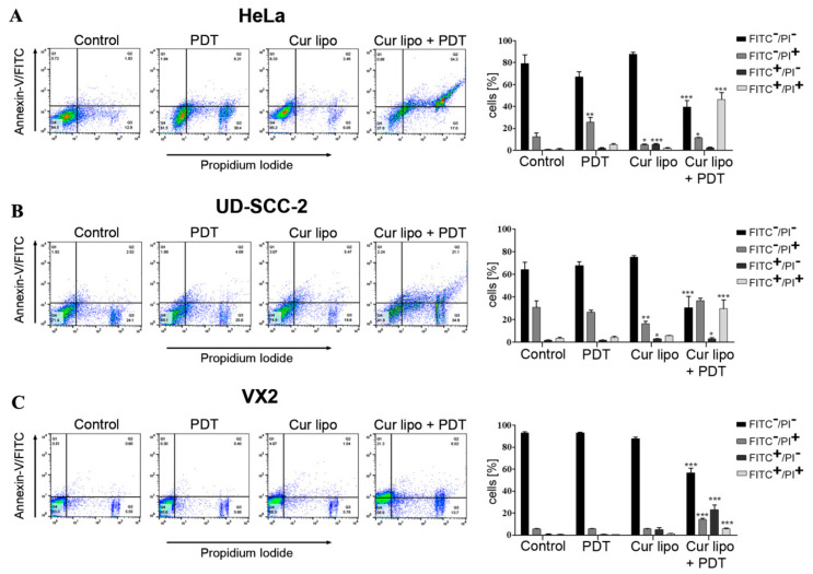
Figure 3.
Evaluation of apoptosis as a cause of cell death in HeLa, UD-SCC-2, and VX2 cells after treatment with curcumin liposomes (Cur lipo) and photodynamic therapy (PDT). All cells were incubated with Cur lipo for 4 h and subsequently irradiated with a light fluence of 3 J·cm−2. After 24 h treatment, cells were co-stained with Annexin V-FITC (fluorescein isothiocyanate) and PI (propidium iodide). Representative flow cytometry micrographs are shown for HeLa (A), UD-SCC-2 (B), and VX2 (C) cells treated with PDT alone, Cur lipo alone, or a combination of Cur lipo with PDT. Untreated cells were used as a control. Q1 represents early apoptotic cells, Q2 represents late apoptotic or necrotic cells, Q3 represents necrotic cells, and Q4 represents live cells. Bar graphs represent the percentage of total apoptotic cells from at least three experiments. Data are shown as mean ± standard deviation, and statistical significances are indicated as *** p < 0.001, ** p < 0.01, * p < 0.05.

