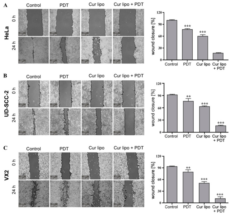
Figure 6.
Cell migration (wound healing) assay in HeLa, UD-SCC-2, and VX2 cells. HeLa (A), UD-SCC-2 (B), and VX2 (C) tumor cells were treated with photodynamic therapy (PDT) only, curcumin liposomes (Cur lipo) only, or Cur lipo in combination with PDT. Images were captured at t = 0 h directly after scratching the cell layer and t = 24 h to evaluate scratch (wound) closure indicative of cellular migration. The cell-free area of the scratched region was measured with the Montpellier Ressources Imagerie (MRI) wound healing tool used with the ImageJ analysis software [41]. The level of cell migration is presented as the percentage of scratch (wound) closure observed 24 h after treatment compared to control values. Controls indicate untreated cells. The values are expressed as the mean ± standard deviation, and statistical significances are indicated as *** p < 0.001, ** p < 0.01.

