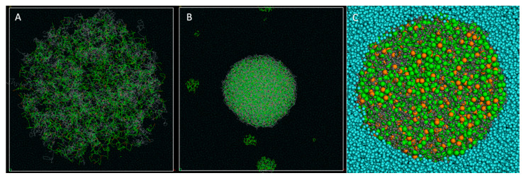
Figure 6.
DPD simulation for the PLA particle formation with vitamin E: (A) Starting point of the DPD simulation. A droplet with a radius of 130 Å containing a mixture of water, PLA and vitamin E molecules (8% loading rate) is placed at the center of a water-filled simulation box. (B) End of the simulation after 30 ns total simulation time. The vitamin E molecule is totally dispersed within the PLA chains, from the surface to the core of the particle. The scale factor is the same as in (A) for better observation of the volume contraction. The whole simulation cell (280 × 280 × 280 Å) is displayed in A and B. (C) Zoomed-in cross-section of the particle. The water molecules are totally excluded from the hydrophobic core formed by the intricated PLA chains and the vitamin E molecules.

