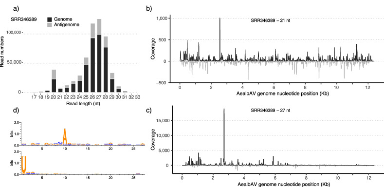
Figure 4.
AealbAV-specific miRNAs in Ae. albopictus samples infected with CHIKV. (a) Size distribution of miRNAs mapping to the AealbAV genome (black) and antigenome (grey). (b) Mapping profiles of the 21-nt reads representing AealbAV-derived siRNAs and (c) the 27-nt reads representing AealbAV-derived piRNAs. (d) Relative nucleotide frequency and conservation of the 27-nt vpiRNAs that mapped to the genome (upper panel) and the antigenome (bottom panel) of AealbAV. AealbAV-derived vpiRNAs display the characteristic piRNA ping-pong signature with Adenine at position 10 for the genomic strand and Uridine at position 1 for the antigenomic strand.

