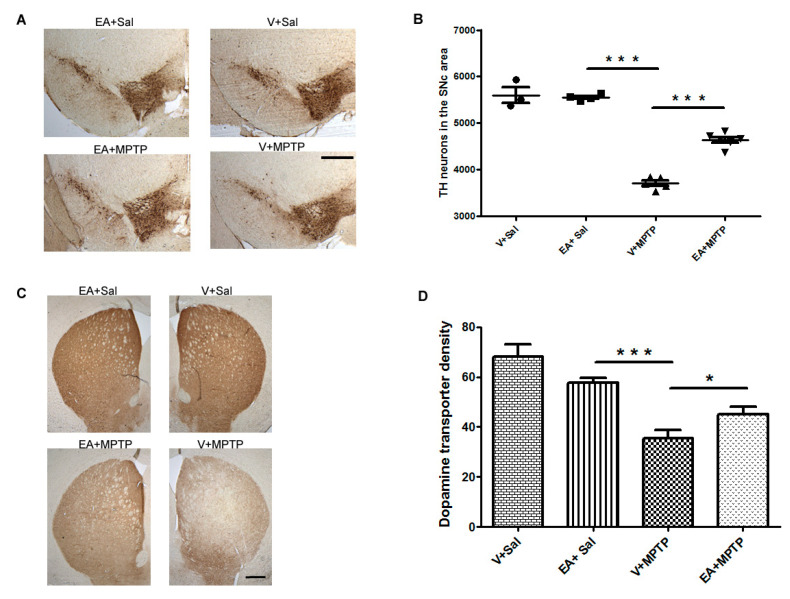
Figure 1.
Immunostaining of tyrosine hydroxylase immune-positive (TH+) neurons to quantify the number of dopaminergic (DA) neurons in the substantia nigra (SNc) and dopamine transporter (DAT) in the striatum. (A): Representative images showing the TH+ neurons in the SNc area. (B): The number of DA neurons in the SNc was counted in each animal using unbiased Stereo Investigator system as described in Section 2. The number of DA neurons was significantly higher in the SNc of the control group (V + Sal) when compared to the MPTP-injected group (V + 1-methyl-4-phenyl 1,2,3,6 tetrahydropyridine (MPTP)). The ellagic acid (EA) treatment significantly prevented the DA neurons from the MPTP-induced neurodegeneration (n = 3–6 animals). (C): Representative images showing the immunoreactivity of dopamine transporter (DAT) in the striatum. The intensity of dopamine nerve terminals was significantly reduced in the striatum of MPTP-injected mice when compared with that in the control (V + Sal) group. Treatment with EA prior to MPTP injection shows significant attenuation of dopamine nerve terminals intensity. (D): DAT intensity was measured using NIH image J software and is presented in the graph. Values are expressed as a percentage of mean ± SEM relative to 100% control (n = 3–5 animals). The scale bar is 400 µm. Significance is denoted as * p < 0.05 and *** p < 0.001.

