Figure 1(A and B).

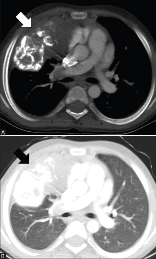
CT images of a 10-year-old male with IMT. Axial images in mediastinal and lung window (A and B) show a large soft tissue mass with extensive calcifications in the right lung (arrows) with possible mediastinal invasion

CT images of a 10-year-old male with IMT. Axial images in mediastinal and lung window (A and B) show a large soft tissue mass with extensive calcifications in the right lung (arrows) with possible mediastinal invasion