
Figure 2(A and B).
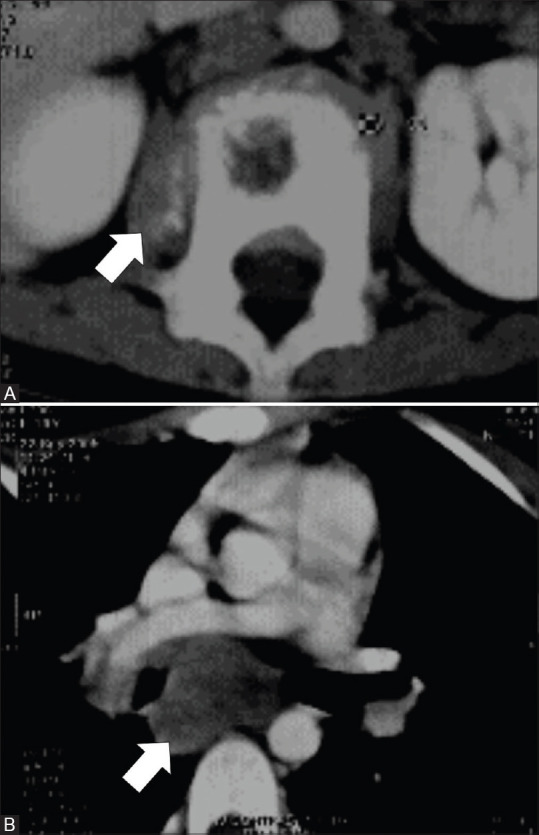
CT images of a 10-year-old female with presumed multifocal IMT of the mediastinal lymph nodes and L1 vertebra. Axial image (A) show lytic lesion in L1 vertebra with adjacent paravertebral soft tissue component showing foci of calcifications (arrow). Axial image (B) show subcarinal lymph nodal mass without calcification (arrow)