
Figure 3(A and B).
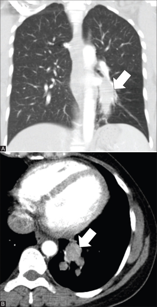
CT images of a 37-year-old female with endobronchial IMT. Coronal image in lung window (A) and axial image in mediastinal window (B) show an oblong and branching, mildly enhancing 6.3 × 2.3 cm, endobronchial mass (arrows) in the posterior basal segmental and subsegmental bronchi of left lower lobe