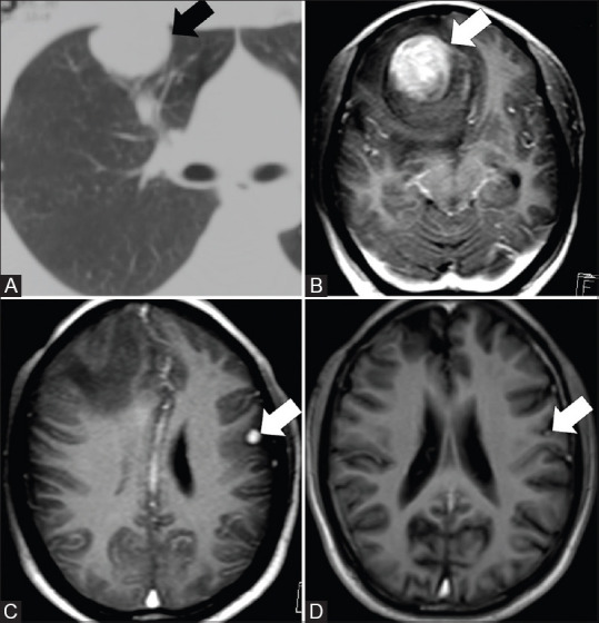
Figure 4(A-D).

CT thorax and MRI brain images of a 35-year-old male with metastases to the brain after 36 months of surgical wedge resection of IMT in the right upper lobe. Axial CT thorax (A) shows the right lung mass (arrow). Axial post contrast MRI brain images (B and C) done a few years after resection of IMT in lung showed a dominant mass in the right frontal region (arrow) and another in the left parietal region (arrow). Axial post contrast MRI brain after radiation showed reduction in size (arrow) and number of lesions (D)
