
Figure 5(A-F).
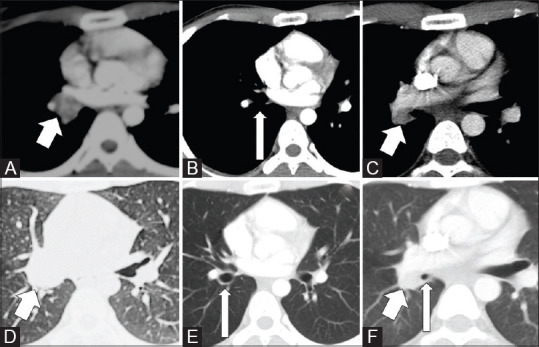
CT images of a 20-year-old male with recurrence of endobronchial IMT after 20 months of bronchoscopic therapeutic removal of tumour. Axial images in mediastinal (A) and lung window (D) shows the endobronchial mass (arrows) and axial images in mediastinal (B) and lung window (E) after endoscopic removal of tumour shows clear bronchus (arrows). CT thorax images after 20 months (C and F) showed recurrent tumour (short arrows) with narrowing of the bronchus (long arrow)