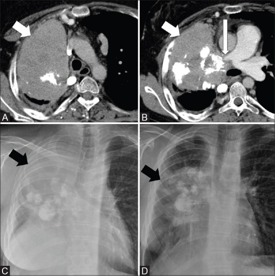
Figure 6(A-D).

CT images and CXR of a 23-year-old male with malignant transformation of IMT to sarcoma after 9.5 years of initial diagnosis. Axial CT thorax images (A and B) shows a large right lung mass with chunky calcifications (short arrows) infiltrating the right pulmonary artery (long arrow). CXR taken after 9.5 years showed considerable increase in size of the mass (arrow) (C) as compared to mass (arrow) at initial presentation (D). Note the elevated right hemidiaphragm in C, which is likely due to combination of infiltration of right main bronchus and phrenic nerve involvement
