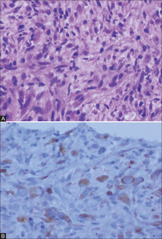
Figure 7(A and B).

Photomicrographs of histopathology and immunohistochemical staining for ALK-1 in inflammatory myofibroblastic tumour. Spindle cells with mild pleomorphism admixed with lymphocytes and plasma cells in a collagenised background (H and E 200×) (A). Granular cytoplasmic staining for ALK-1 in the spindle cells (200×) (B)
