
Figure 13(A-D).
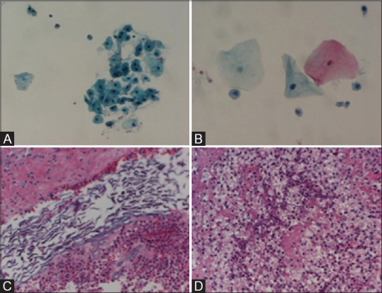
Case 3. (A) FNA, filter preparation, Papanicolaou stain, 200× magnification, mature squamous cells, and scant neutrophils. (B) FNA, filter preparation, Papanicolaou stain, 400× magnification, mature squamous cells, and scant neutrophils. (C) Biopsy, H and E stain, 200× magnification, keratinous material and acute inflammatory infiltrate. (D) Biopsy, H and E stain, 200× magnification, and mixed inflammatory infiltrate with abundant foamy macrophages