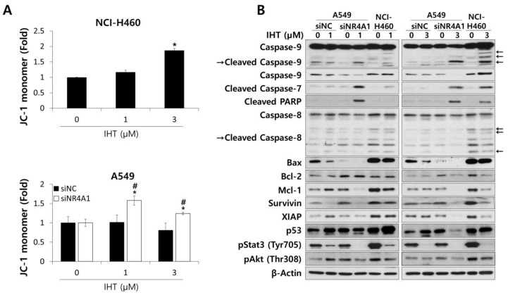
Figure 5.
IHT induced mitochondria-mediated apoptosis in 3D-cultured NCI-H460 cells and A549 cells treated with siNR4A1. (A) To detect alteration of mitochondria membrane potential, tumorspheroids were dissociated into single cells after IHT treatment for 48 h. Cells were stained with tetraethylbenzimidazolylcarbocyanine iodide (JC-1) and analyzed by flow cytometry. *, significantly different from siNR4A1 control (p < 0.05); #, significantly different from siNC control (p < 0.01). All data are presented as mean ± SD. (B) Whole cell lysates were prepared from NCI-H460 and siRNA-transfected A549 tumorspheroids 48 h after IHT treatment. The levels of apoptosis-related proteins were analyzed by western blotting. β-Actin was used as a loading control.

