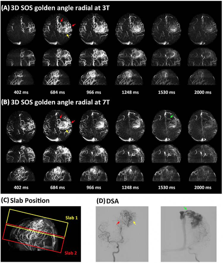
Figure 7.
An AVM case with two slabs acquired with 4D MRA using golden-angle stack-of-stars radial acquisition at 3T (A) and 7T (B). Six representative maximum intensity projection (MIP) images are displayed along axial, coronal, and sagittal views. The positions of the two slabs are shown in the TOF image (C). DSA images serve as the gold standard (D). The entire AVM lesion including feeding arteries (red arrow), nidus (yellow arrow), and draining vein (green arrow) was captured in a two-slab radial 4D MRA. 4D MRA matches well with DSA. It can be noted that the draining vein (green arrow in b) is better depicted at 7T than that at 3T. Adapted from Cong F, Zhuo Y, Yu S, et al. Noncontrast-enhanced time-resolved 4D dynamic intracranial MR angiography at 7T: A feasibility study. J Magn Reson Imaging. 2018;48(1):111–120; with permission.

