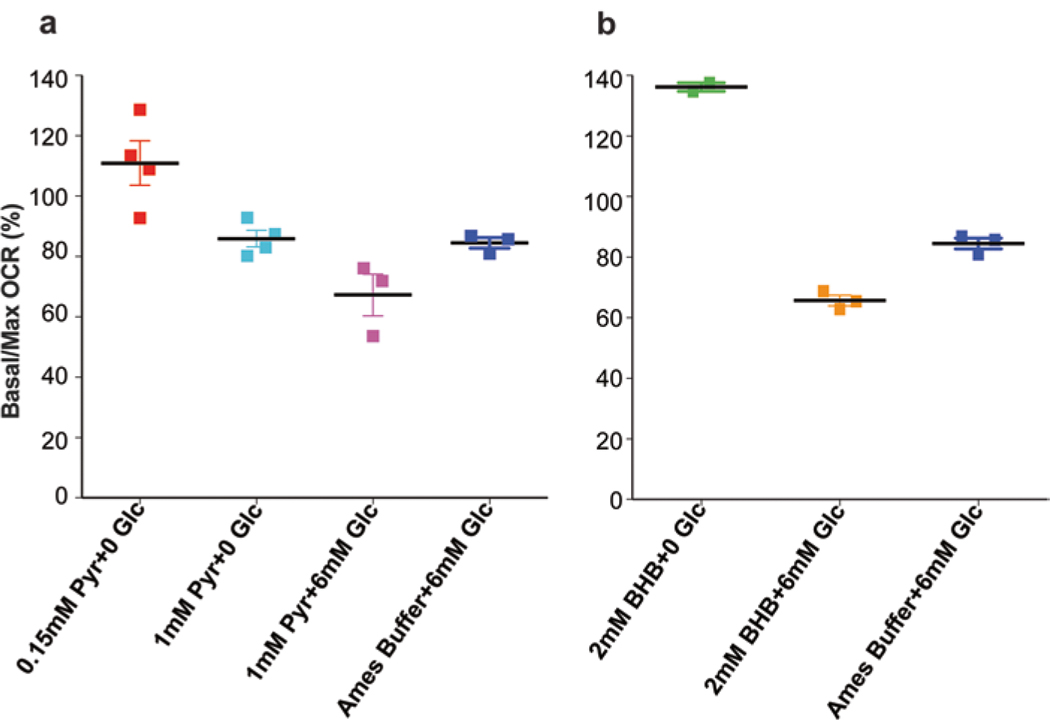
Fig. 3.
Different substrates reveal differences in reserve capacity. (a) Pyruvate was used as a substrate at 0.15 mM and 1 mM concentration with or without glucose. (b) β-hydroxybutyrate was used at 2 mM concentration with or without glucose. Optimal reserve capacity was obtained after the addition of glucose to either substrate. Adult mouse retina was used for all experiments

