Fig. 4.
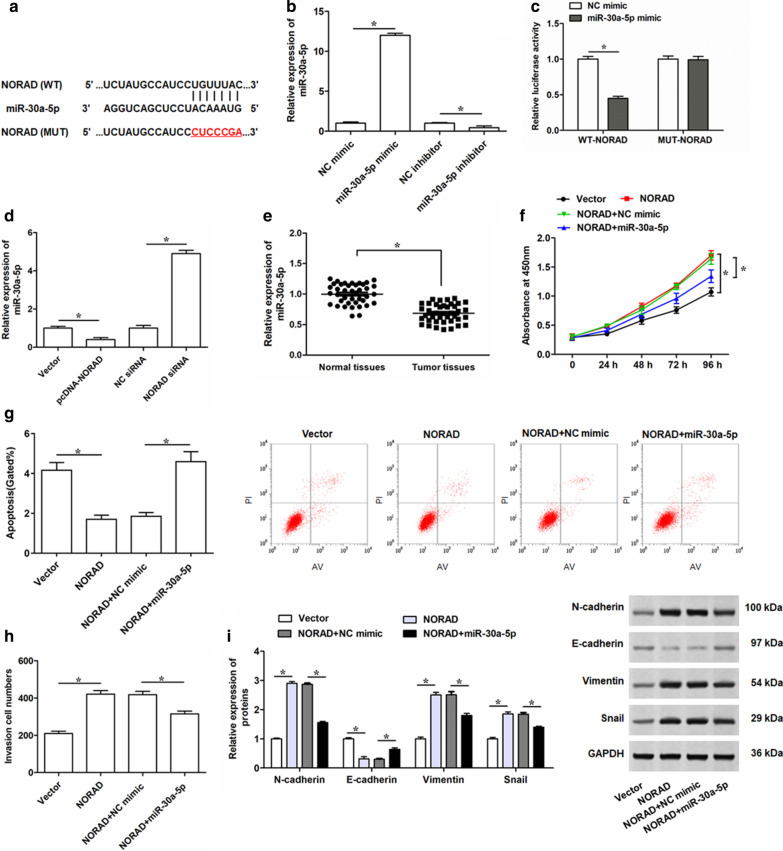
Reintroduction of miR-30a-5p weakens the effect of NORAD overexpression on cell proliferation, invasion, apoptosis and EMT process of PC-3 cells. a The potential binding sequences between NORAD and miR-30a-5p predicted by starBase. b qRT-PCR was conducted to determine the expression levels of miR-30a-5p in PC-3 cells transfected with 100 nM miR-30a-5p mimic, 100 nM miR-30a-5p inhibitor, or 100 nM their negative controls for 48 h. c Dual luciferase reporter assay was performed to determine the luciferase activity of HEK 293 T cells transfected with miR-30a-5p mimic and luciferase reporter vectors containing WT- or MUT- NORAD segment. d The expression level of miR-30a-5p was detected by qRT-PCR in PC-3 cell after being transfected with 2 μg/mL pcDNA-NORAD, 30 nM NORAD siRNA, or their negative controls for 48 h. e qRT-PCR was performed to determine the expression levels of miR-30a-5p in PC tissues (Tumor tissues, n = 45) and adjacent normal tissues (Normal tissues, n = 45) determined by qRT-PCR. Then, PC-3 cells were transfected with 2 μg/mL pcDNA-NORAD alone, or together with 100 nM miR-30a-5p mimic. f Cell proliferation of PC-3 cells after infection for 24, 48, 72 and 96 h were detected by CCK-8 assay. g, h After 48 h transfection, cell apoptosis and invasion was determined by Flow cytometry and Transwell, respectively. i The expression levels of N-cadherin, E-cadherin, Vimentin and Snail in PC-3 cells after infection of 48 h were detected by Western blotting. The data were presented as the mean ± standard error of mean (SEM). Student’s t-test was used for the comparison between 2 groups in this study. *P < 0.05

