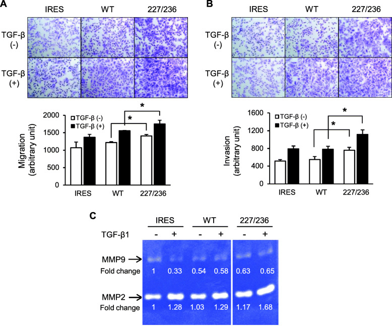
Fig. 3.
Migratory and invasive capabilities of stable HSC-2 cells expressing exogenous TβRII. a and b. Stable transfectant cells harboring an empty vector (IRES), wild-type TβRII (WT), or I227T/N236D TβRII (227/236) were seeded in a transwell chamber (a) or transwell chamber precoated with Matrigel (b). Cells that migrated to the other side of the chamber were counted after 24 h of incubation in the presence of vehicle (TGF-β1 (−)) or 10 ng/ml of TGF-β1 (TGF-β1 (+)); magnification, × 100. Data represent mean ± standard deviation (*P < 0.05). c. Gelatinolytic activities of MMP-2 and MMP-9 were assayed. Stable transfectant cells were incubated in P medium containing 0.2% FBS in the presence of vehicle (−) or 10 ng/ml of TGF-β1 (+) for 24 h. The conditioned medium was collected and subjected to gelatin zymography. Samples of IRES, WT, and 227/236 were assayed on the same gel and the images were cropped. IRES, empty vector; WT, wild-type TβRII; 227/236, I227T/N236D TβRII. Uncropped gelatin zymography was shown in Additional file 4: Figure S4

