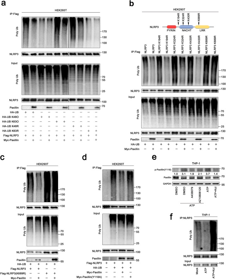
Fig. 5.
Paxillin promotes NLRP3 K63-linked deubiquitination depending on extracellular ATP and K+ efflux. a HEK293T cells were co-transfected with pFlag-NLRP3 and pHA-UB, pHA-UB(K48O), pHA-UB(K63O), pHA-UB(K48R), pHA-UB(K63R), or pMyc-Paxillin. b HEK293T cells were co-transfected with pHA-UB and pFlag-NLRP3, pFlag-NLRP3(K194R), pFlag-NLRP3(K324R), pFlag-NLRP3(K403R), pFlag-NLRP3(K689R), or pMyc-Paxillin. c HEK293T cells were co-transfected with pHA-UB and pFlag-NLRP3, pFlag-NLRP3(K689R), or pMyc-Paxillin. d HEK293T cells were co-transfected with pFlag-NLRP3 and pHA-UB, pMyc-Paxillin, or pMyc-Paxillin(Y118A). e TPA-differentiated THP-1 macrophages were treated with DMSO, A438079 (10 μM), or AZ10606120 (10 μM) for 1 h before the treatment with ATP (5 mM) for 2 h or treated with ATP (5 mM) for 2 h and with/without 50 mM extracellular KCl. The protein levels of p-Paxillin-(y118), Paxillin, and GAPDH were determined by Western blotting. f TPA-differentiated THP-1 macrophages were treated with ATP (5 mM) for 2 h in the presence or absence of 50 mM extracellular KCl. Lysates were prepared and subjected to denature-IP (top) or subjected to Western blot (as input) (bottom) (a–m). Densitometry of the blots was measured by ImageJ

