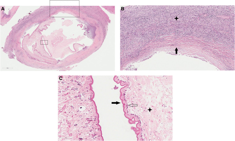
Figure 2.
Histopathology of the excised cystic lesion. The cyst and adjacent brain tissue stained with hematoxylin and eosin. (A) Cysticercus (×40 magnification) with surrounding fibrous capsule and inflamed brain parenchyma. (B) Fibrous capsule (black arrow; ×100 magnification) and adjacent white matter inflammatory cell infiltrate (star) consisting of lymphocytes, plasma cells, histiocytes, and eosinophils. (C) Cyst wall tegument (×400) demonstrating outer microvilli (black arrow), cellular layer (transparent arrow), and inner reticular layer with excretory canaliculi (star). This figure appears in color at www.ajtmh.org.

