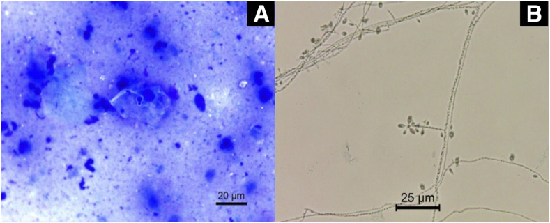
Figure 1.
(A) Giemsa-stained direct microscopy showing the presence of rare and budding yeast cells (×630). (B) Micromorphology from the colony grown on potato dextrose agar for 7 days at 25°C showing septate mycelial filaments, delicate conidiophores, and conidia arranged sympodially with floral appearance. Lactophenol blue cotton staining (×630). This figure appears in color at www.ajtmh.org.

