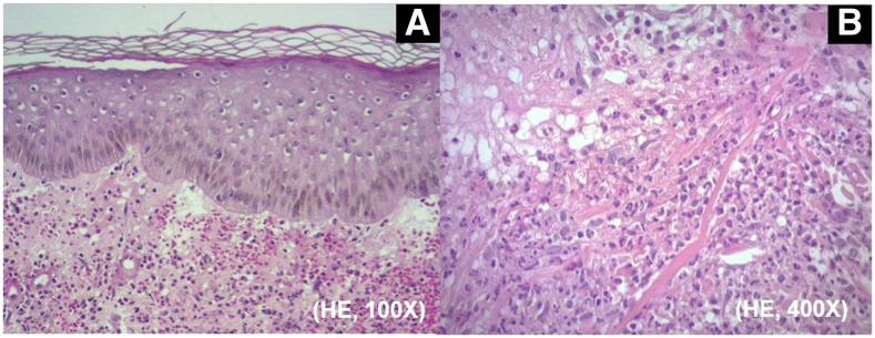
Figure 2.
(A) Histopathology of an erythematous cutaneous lesion from patient 6, compatible with Sweet syndrome. Diffuse inflammatory infiltrates associated with papillary dermal edema. (B) Close-up view of the infiltrates composed predominantly of neutrophils with leukocytoclasia (hematoxylin and eosin; original magnification: [A] ×100; [B] ×400). This figure appears in color at www.ajtmh.org.

