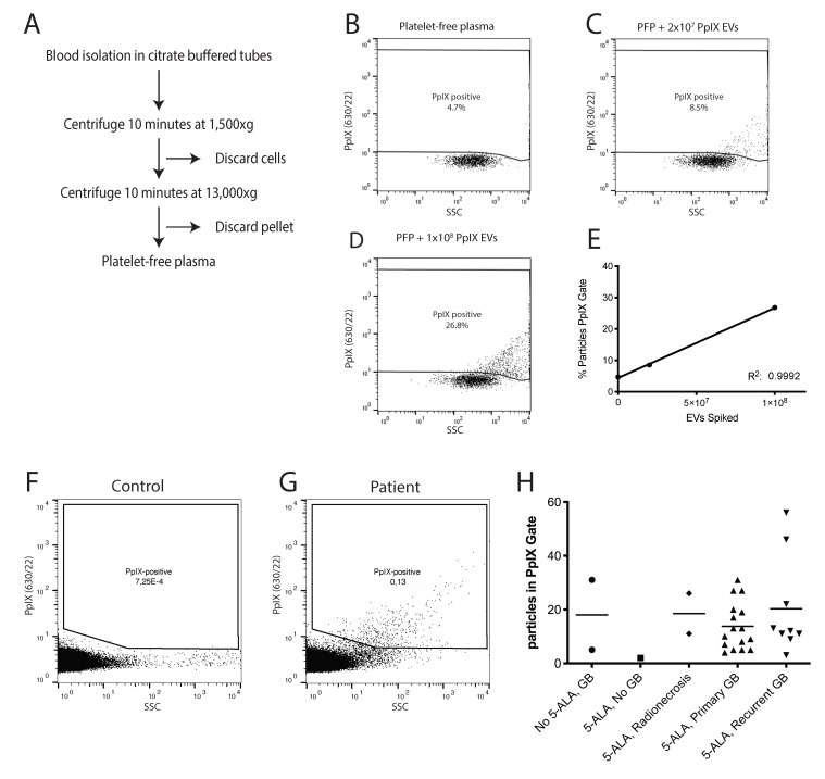
Figure 2.
Isolation of PpIX-positive EVs from patient plasma. (A) Centrifugation protocol for the isolation of platelet-free plasma from whole blood. (B) hFC analysis of platelet-free plasma. (C) hFC analysis of platelet-free plasma spiked with 2 × 10⁷ U87 derived PpIX-positive EVs. (D) hFC analysis of platelet-free plasma spiked with 1 × 10⁸ U87 derived PpIX-positive EVs. (E) Linear regression plot of the % of particles in the PpIX gate versus the number of PpIX EVs spiked in. Goodness of fit, R2: 0.9998. (F) hFC analysis of PpIX particles in platelet-free plasma from control (10 min measurement). (G) hFC analysis of PpIX particles in platelet-free plasma from a patient treated with 5-ALA (10 min measurement). (H) Number of particles in the PpIX gate per patient group detected during 5 min of measurement.

