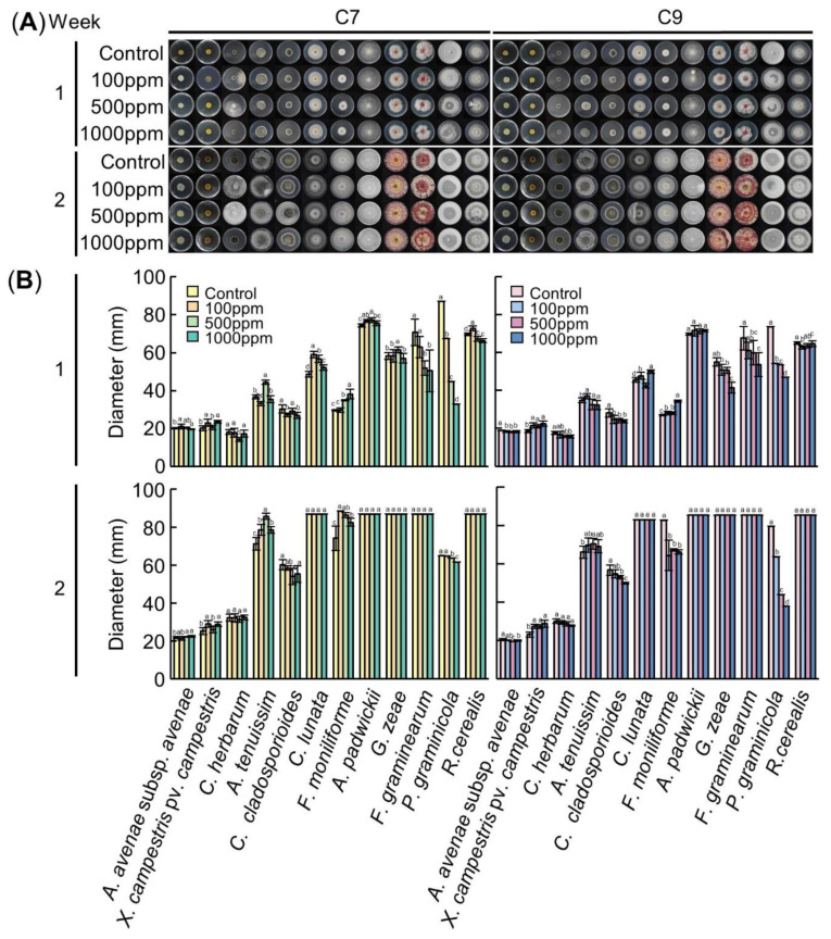
Figure 3.
Antimicrobial activity of C7 (left panel) and C9 (right panel). (A)Visual growth of different bacterial and fungal strains. Bacteria were cultured in an LB medium and fungi were cultured in a PDA medium at 25 °C in a dark state. The medium was treated with C7 and C9 at concentrations of 0 ppm, 100 ppm, 500 ppm and 1000 ppm, respectively. Colonies were observed after 1 and 2 weeks of growth after inoculation. (B) Colony diameter according to the concentration of the compounds at 1 week and 2 weeks after inoculation. Means with the same letters are not significantly different by Duncan’s multiple range test at p < 0.05.

