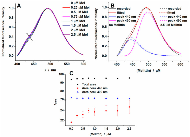
Figure 2.
(A) Fluorescence emission spectra of Laurdan inserted into 1,2-dioleoyl-sn-glycero-3-phosphocholine (DOPC) liposomes (37 °C) at increasing concentrations of melittin (B), deconvoluted spectra for DOPC liposomes alone or in the presence of the highest melittin concentration from (A) and (C) the dependence of the area values vs. melittin concentration.

