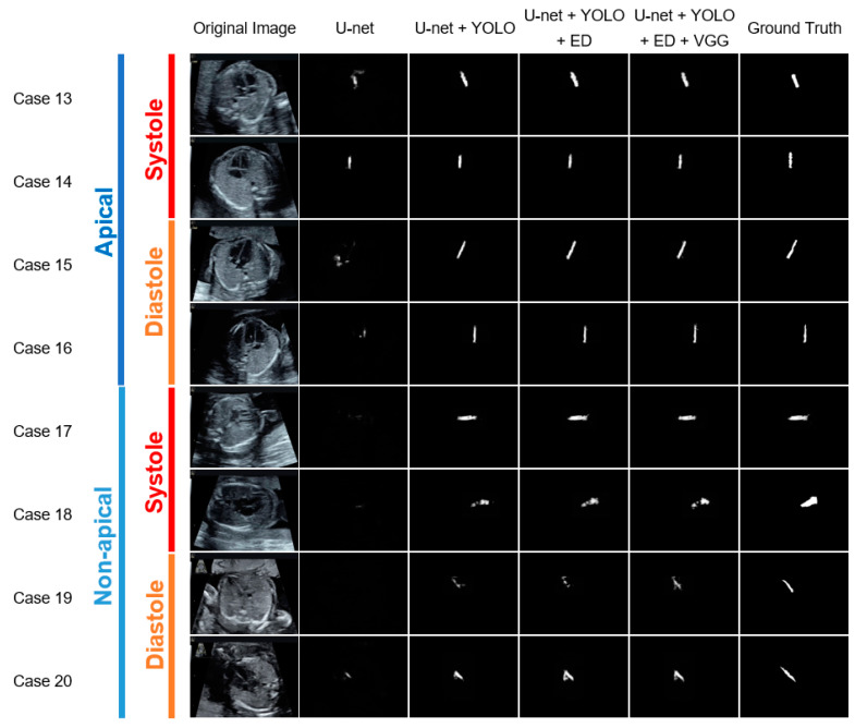
Figure 5.
Representative examples of the ventricular septum segmentation images classified by the cardiac axis orientation and ventricular systolic state, from the test data, for each module combination. One horizontal row presents the segmentation results obtained using each method for the same case. The white pixels are estimated as the ventricular septum, and the whiteness indicates the confidence level. The segmentation results were more accurate for the apical group than for the non-apical group, and more accurate for the diastolic group than for the systolic group, irrespective of the module combination. The addition of the YOLO significantly improved the segmentation accuracy, and the addition of the ED further improved it, irrespective of cardiac axis orientation and ventricular systolic state. Moreover, the addition of VGG slightly improved the segmentation accuracy for the systolic and non-apical groups.

