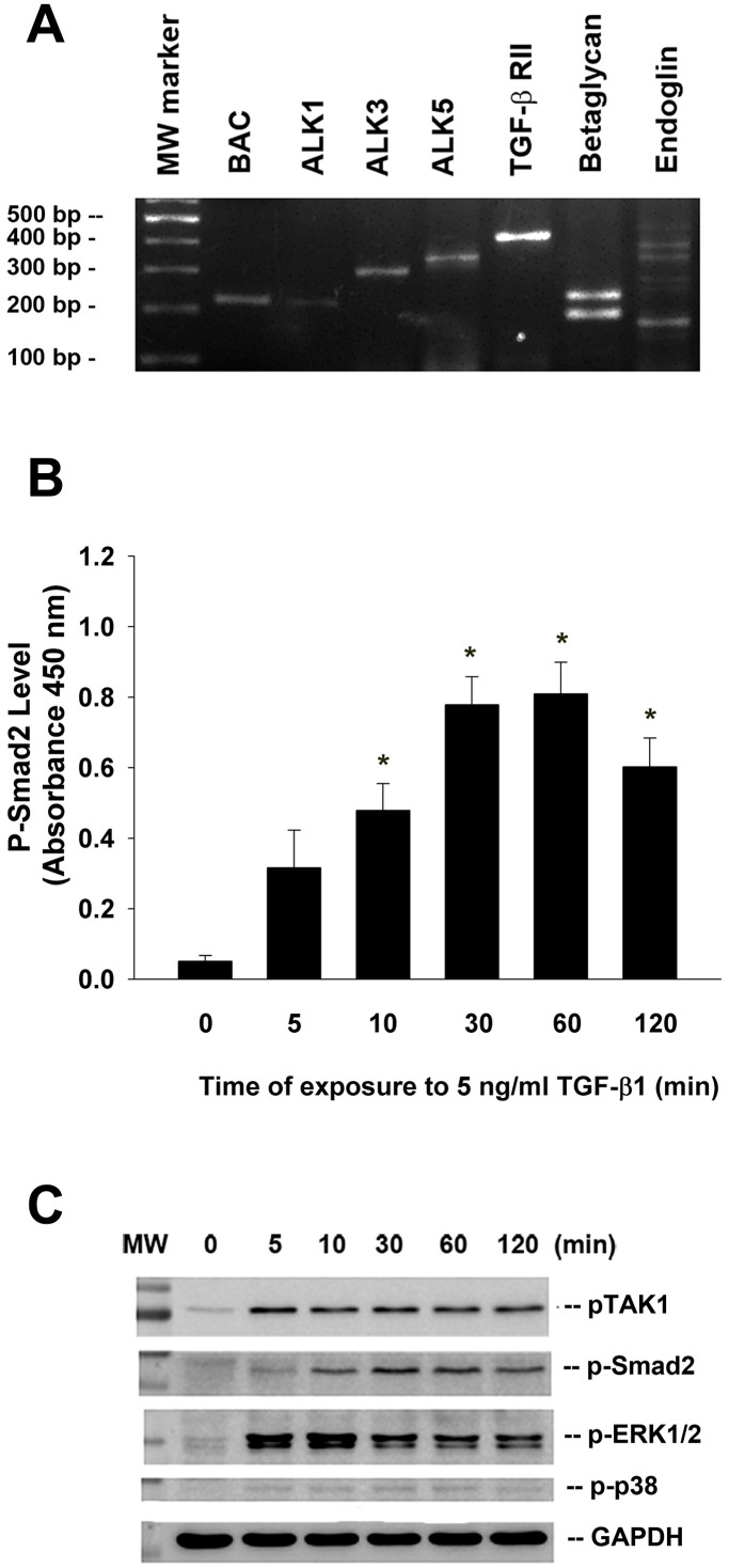
Figure 2.
Expression of various TGF-β related receptors in SHED and the effect of TGF-β1 on the Smad2, TAK1, ERK1/2 and p38 phosphorylation of SHED. (A) SHED cells were cultured in DMEM with 10%FBS for 24 hours. Total RNA was isolated for RT-PCR analysis of TGF-β related receptors (ALK1, ALK3, ALK5, TGF-β1RII, betaglycan, endoglin) expression, (B) SHED were exposed to TGF-β1 for 0-120 min (as indicated on graphs). Cell lysates were prepared and proteins were used for analysis of p-Smad2 expression by PathScan phospho-ELISA (OD450, Mean ± SE). *Denotes statistically significant difference when compared with control. (C) SHED were exposed to TGF-β1 for 0-120 min. Cell lysates were prepared and proteins were used for western blotting analysis of p-Smad2, p-TAK1, p-ERK1/2, p-p38 and GAPDH (control) protein expression. One representative western blotting picture was shown.

