Figure 4.
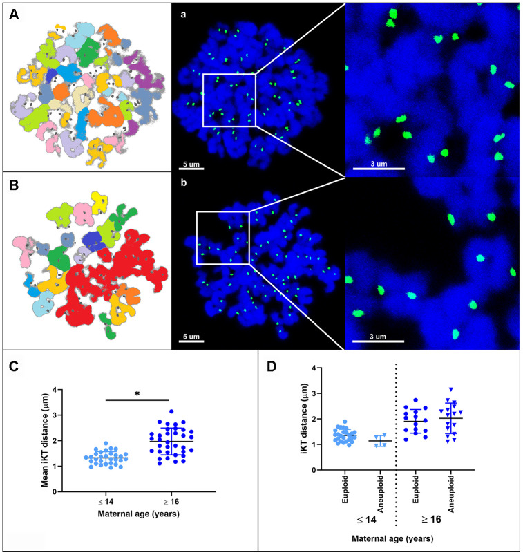
Representative maximum projection images of chromosome spreads for in vitro matured MII oocytes from young (A) and aged (B) mares. Green, kinetochores (CREST); blue, chromatin (Hoechst 33342). (A, B) explanatory drawing of the chromosome spreads. Different sister chromatid pairs have different colors. Note the increased interkinetochore distance (i.e. separation of the CREST signals) in oocytes from old mares. Bar, 5 and 3 μm. Scatterplots of interkinetochore distance categorized by mare age (C) (*, p < 0.0001) and by mare age and aneuploidy (D).

