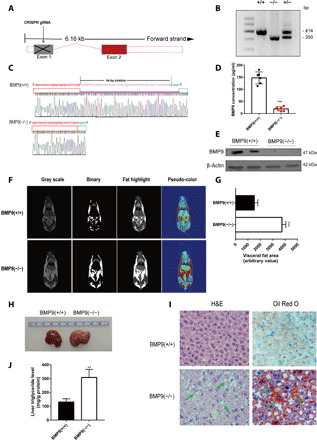
Fig. 2. BMP9-KO mice exhibit a distinct hepatic steatosis phenotype.

(A) Schematic diagram of BMP9 deletion process by targeting exon 1 via CRISPR guide RNA (gRNA). (B) Genotyping of BMP9 WT, KO, and heterozygous mice. (C) Gene sequencing results. (D) BMP9-KO mouse serum BMP9 protein concentrations (n = 6). (E) Hepatic BMP9 protein expression in BMP9-KO mice (n = 2). (F) NMR showed distribution of mice visceral fat. Photo credit: Z. Yang, Photographer Institution, Ninth People’s Hospital, Shanghai Jiao Tong University School of Medicine. (G) Quantification of visceral fat area (n = 3). (H) Representative image of a dysfunctional liver in 16-week-old BMP9 WT and KO mice fed a CD (n = 6). Photo credit: P. Li, Photographer Institution, Ninth People’s Hospital, Shanghai Jiao Tong University School of Medicine. (I) Liver H&E and Oil Red O staining (original magnification, ×400). (J) Liver TG level. **P < 0.01 and ***P < 0.001.
