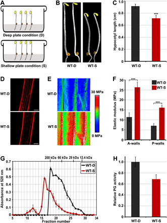
Fig. 1. Pectin degradation is involved in touch-regulated hypocotyl cell elongation and cell wall mechanics.

(A) Schematic diagram of the deep-shallow plate assay. (B and C) Images (B) and hypocotyl length (C) of 3-day-old etiolated seedlings grown in the D or S condition. Mean ± SD; n ≥ 20. (D) PI staining images of cells at the middle region of hypocotyls. Scale bar, 100 μm. (E and F) Elastic modulus map (E) and quantification (F) of epidermal cell walls at the middle region of hypocotyls. The elastic modulus on the A-walls and P-walls was obtained by AFM in the QNM mode. Mean ± SD; n ≥ 10. (G) Molecular mass profile of CDTA-soluble pectin in 3-day-old etiolated seedlings grown in the D or S condition. The mean ± SD was obtained from two technical replicates. (H) In vivo PG activity of etiolated seedlings in response to mechanical resistance stimulation. The relative PG activities of the samples were normalized to the absorbance of wild-type (WT) seedlings grown in the D condition. Mean ± SD; n = 3.
