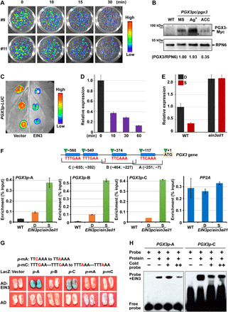
Fig. 4. EIN3 directly binds to the promoter of PGX3 and represses PGX3 gene expression.

(A) Bioluminescent luciferase images showing the gene expression levels of PGX3 with 10-ppm ethylene treatment for the indicated periods. (B) Immunoblot analysis of PGX3 protein levels. PGX3/RPN6 indicates the relative band intensities of PGX3-Myc normalized to RPN6 and is presented relative to that grown on 1/2 MS medium set at unity. (C) Luciferase bioluminescence images of PGX3p-LUC transiently coexpressed with the 35S vector (Vector, left) or 35S-EIN3 (EIN3, right) in tobacco leaves. (D) RT-qPCR analysis of regulation of PGX3 gene expression by β-estradiol–induced EIN3 proteins. Mean ± SD; n = 3. (E) RT-qPCR analysis of PGX3 gene expression in response to mechanical resistance. Mean ± SD; n = 3. (F) ChIP-qPCR analysis of the interactions between EIN3 protein and PGX3 promoter. The top diagram shows the PGX3 promoter fragments, which contain putative EBSs (blue rectangles). Mean ± SD; n = 3. (G) Yeast one-hybrid assay showing that specific elements are required for physical binding of EIN3 to PGX3 promoter. The top diagram shows mutated PGX3 promoter fragments A and C with mutations located in the EBSs. (H) EMSA indicating physical binding between EIN3 and PGX3 promoter.
